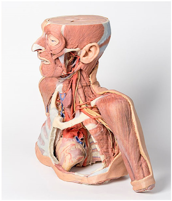
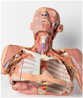
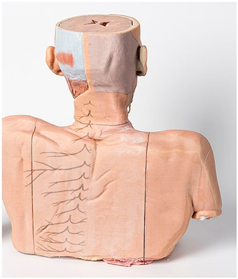
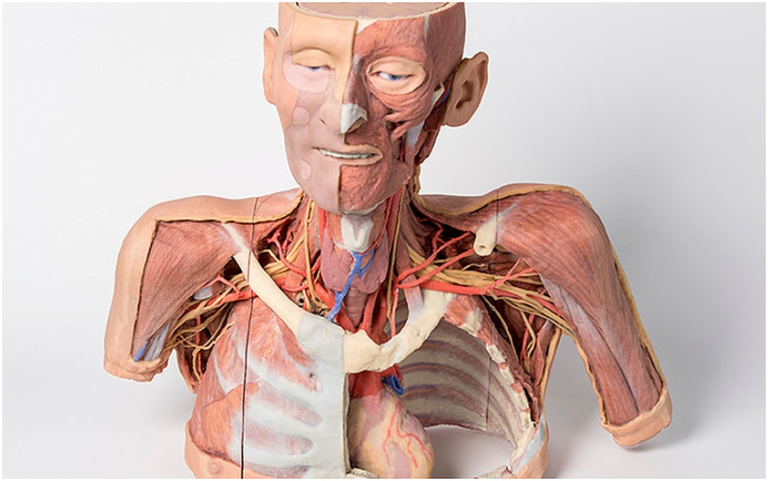
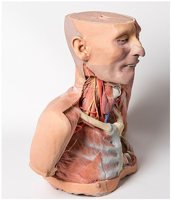
Head and neck: The head and neck of the specimen provides views of both superficial and deep structures in the region. The calotte has been removed ~2cm superior to the orbits to expose the brain in relation to the endocranial cavity. The transverse section through the cerebrum demonstrates the relation of the grey matter cortex to the white matter medulla, as well as the lateral ventricles with a small amount of choroid plexus visible in the base of both spaces. The skin and superficial fascia on the right side has been retained and false-coloured to display the angiosomes of the face and posterior neck. On the left side, the superficial tissues have been dissected to expose the muscles of facial expression, muscles of mastication, and deeper structures of the infratemporal fossa including the lingual nerve, terminal branches of the external carotid artery into the superficial temporal and maxillary arteries.
The carotid sheath has been opened on both sides of the neck, and the internal jugular veins and sternocleidomastoid muscles largely removed, to expose the pathway of the common carotid arteries, internal and external carotid arteries, and the vagus nerves. On the right side, the great auricular nerve ascends towards the face, while the hypoglossal nerve can be seen adjacent to the exposed stylohyoid ligament and supra- and infrahyoid muscles. A large thyroid gland is present bilaterally inferior to the thyroid cartilage, with a well-preserved superior thyroid artery and inferior thyroid vein on the right side and across the midline.
The root of the neck – axillary junction: The clavicle has been partially removed on the left side of the specimen (medial to the origin of the deltoid) to expose the first rib and the insertion of anterior scalene muscle. The roots of the brachial plexus (C5-T1) can be seen forming the trunks posterior to this muscle but anterior to middle and posterior scalene muscles they emerge from the interscalene plane. While the subclavian vein has been removed, the subclavian artery is also seen passing behind the scalenus anterior. The transition of the subclavian artery to the axillary artery is exposed, as is its position relative to the cords of the brachial plexus (medial, lateral and posterior).
The left axilla has been dissected to expose the divisions and cords of the brachial plexus and its major and minor branches. The contributions from the medial and lateral cords coming together around the axillary artery to form the median nerve is very distinctive. The course of the medial cord, the ulnar nerve, is clearly visible as is the musculocutaneous nerve as the continuation of the lateral cord. The axillary nerve is seen wrapping posteriorly around the surgical neck of the humerus. The thoracodorsal nerve and artery are seen descending on the medial wall of the axilla to enter the latissimus dorsi muscle. The long thoracic nerve is seen just anterior to this upon the serratus anterior muscle which it supplies.
The axilla/root of neck junction on the right is similar except the clavicle (and subclavius muscle) has been retained, which gives an appreciation of the dimensions of the cervico-axillary canal through which structures gain entry to the axilla. Also on the right side the pectoralis minor and major (that comprise the anterior axillary wall) have been reflected with only a small portion of their insertions being retained.
Thorax: The thorax has been opened via a ‘window’ on the left to display the internal thoracic wall and mediastinum. The left lung has been removed and the intercostal spaces are discernable deep to the parietal pleura although intercostal neurovascular bundles are only discernable posteriorly. The pericardium has been removed to expose the heart with its apex pointing inferiorly, anteriorly, and to the left. The left side of the heart is exposed as are the left pulmonary veins and arteries (above left main bronchus), ascending aorta, aortic arch and commencement of the descending thoracic aorta. The left vagus nerve and left recurrent laryngeal nerve are easily identified. The right half of the anterior and lateral thoracic wall are intact and display the muscles of the intercostal spaces and inserting hypaxial muscles from the right upper limb. If the specimen is viewed from below, the right lung and pleural spaces along with the diaphragmatic surface of the heart are all evident. While the skin and superficial fascia posterior thorax has been left intact, the distribution of cutaneous branches of dorsal rami have been illustrated along the left side of the specimen.


 보건교육지원센터를 시작페이지로!
  즐겨찾기
즐겨찾기


